

Histology and cytology
-
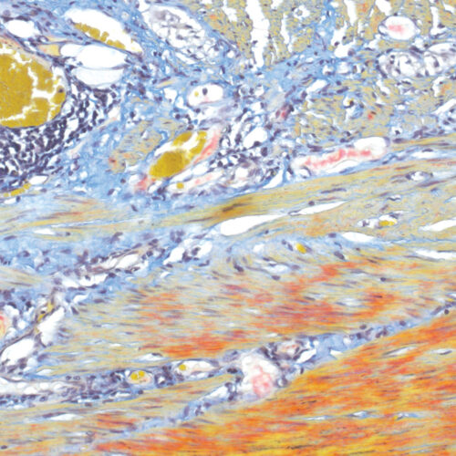
Martius Scarlet Blue (MSB) kit
Seven-reagent kit used for fibrin visualization, especially of older clusters. This method is a modification of Masson Trichrome and is ideal for studying connective tissue and vascular pathology.
-

Martius Yellow C.I. 10315
Golden Yellow, Martinsgelb, Acid yellow 24. For staining erythrocytes yellow in trichrome staining methods.
-
-
Masson Fontana kit
Six-reagent melanin and argentaffin granule staining kit, based on the reduction of silver nitrate to elemental silver. Melanin is a brown-black pigment normally present in the hair, skin, retina, iris and certain parts of CNS. Argentaffin granules are found in carcinoid tumors.
-
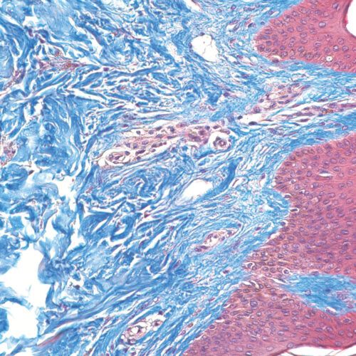
Masson Trichrome kit
Seven-reagent kit for staining muscle and collagen fibers with a blue counterstain. It is also used for visualizing gametes, nuclei, neurofibrils, glial cells, keratins and intercellular fibrils. The kit may be useful for detecting collagen in smooth muscle cancer or diseases like cirrhosis.
-
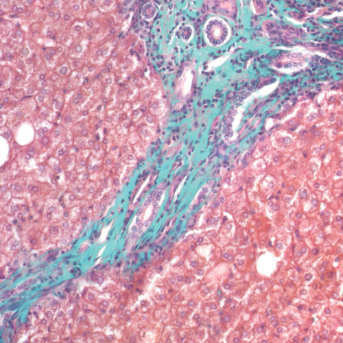
Masson-Goldner Trichrome kit
Seven-reagent kit for staining muscle and collagen fibers with green counterstain. It is also used for visualizing gametes, nuclei, neurofibrils, glial cells, keratins, intercellular fibrils and for differentiation of smooth muscle fibers and collagens.
-
-
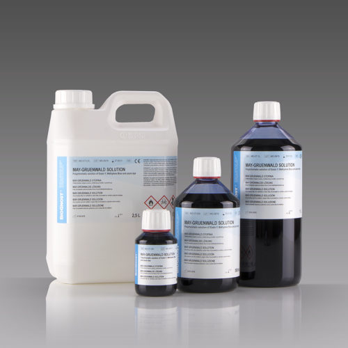 Histology and cytology/Citology and hematology staining reagents/Rapid and standard hematology reagents and kits/May-Gruenwald and Giemsa reagents
May-Gruenwald solution
Polychromatic solution of Eosin Y, Methylene Blue and azure dyes. For staining in hematology, cytology and staining sections of hematopoietic organs in histopathology.